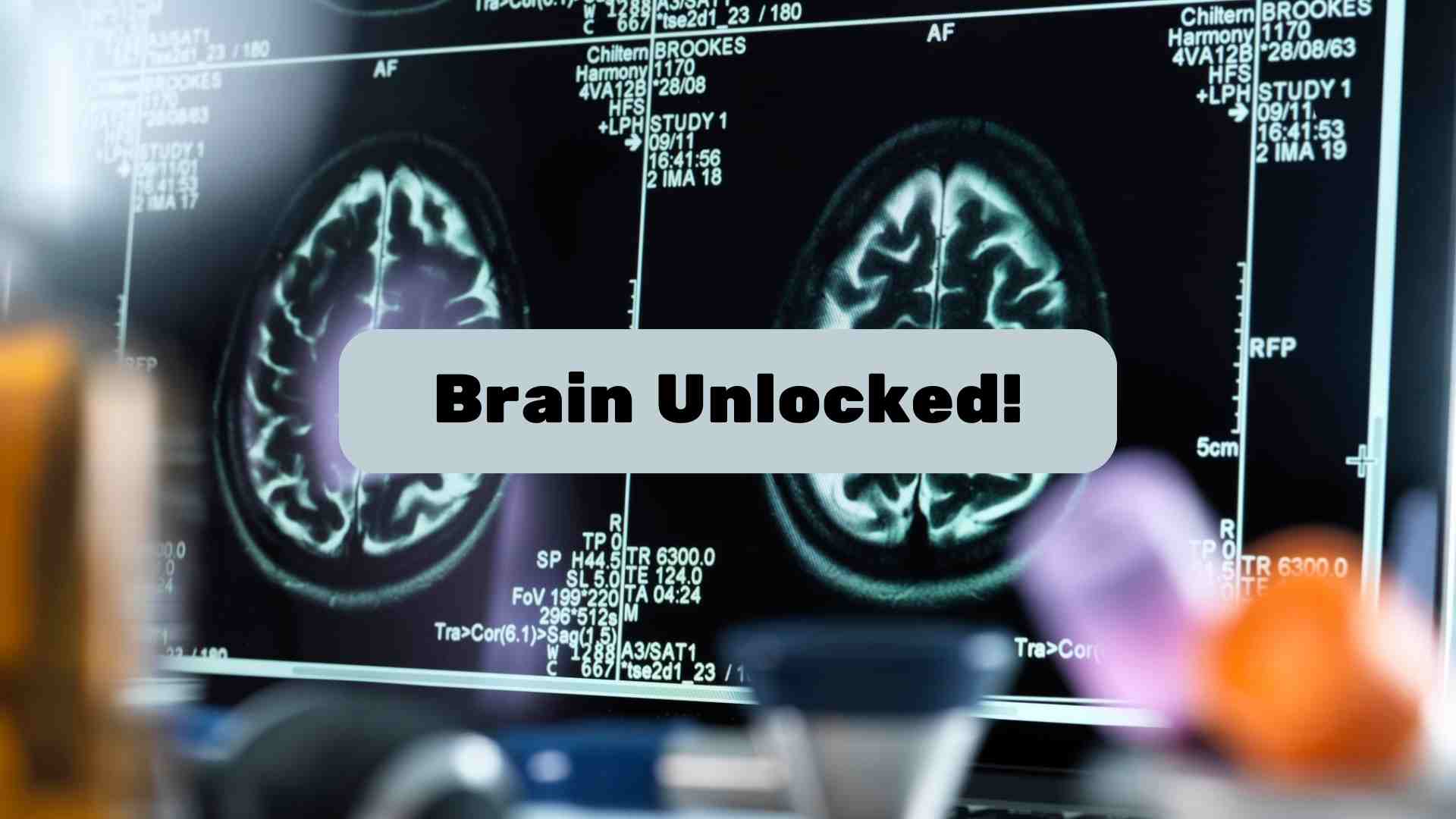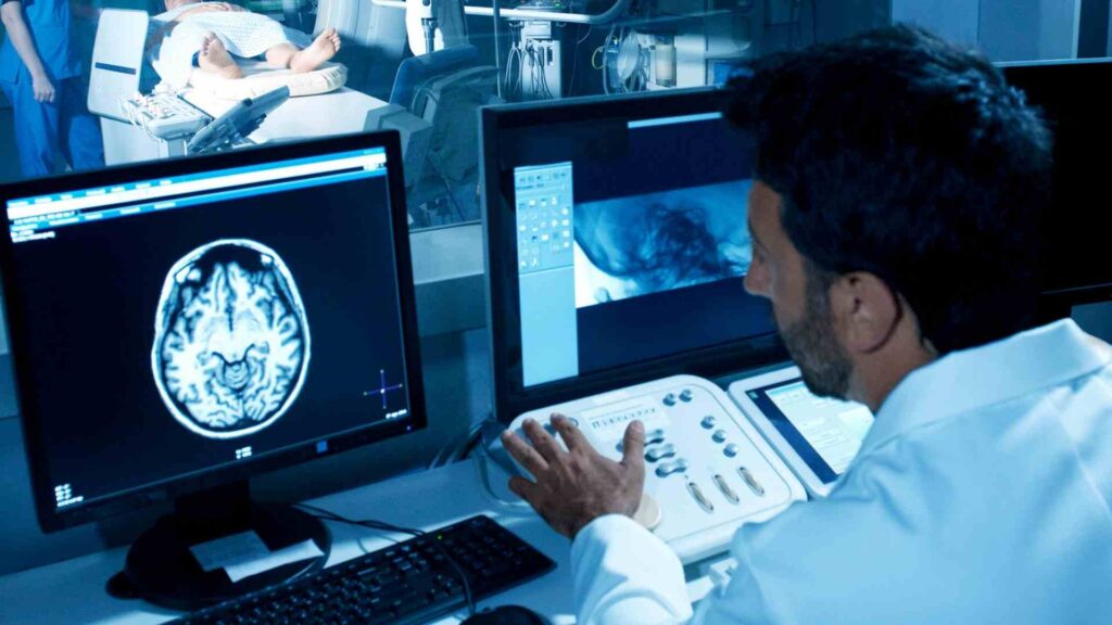
Neuroimaging is a branch of medical imaging sciences that focuses on capturing detailed images of the brain and CNS (Central Nervous System) without the use of invasive methods like surgery. It is also considered similar to the field of modern neuroscience. Neuroimaging includes a variety of quantitative (measurable) techniques that allow one to study the structure and function of the brain in detail. Each technique is designed to reveal different aspects of the brain’s structure and function.
The significance of neuroimaging of the brain can be seen from the fact that it provides a range of directly or indirectly derived visual representations as well as quantitative analysis of the anatomy (internal structure), blood flow, electrical activity, metabolism, oxygen consumption, receptor sites, and many other physiological functions within the CNS. It is important within psychology as well as it allows in-depth study of what certain areas of the brain are responsible for, as well as being able to identify brain differences in those who may have psychological disorders or other kinds of mental health conditions.
These imaging methods have revolutionized our understanding of the brain, aiding in the diagnosis of diseases, the assessment of brain health, and the exploration of how various activities impact brain function. So, the question is how does neuroimaging work? Let’s know more!
A Beginner’s Guide to Neuroimaging Techniques
What is neuroimaging? Neuroimaging of the brain offers a window into the living brain, enabling us to visualize its structure and function non-invasively. It involves the use of various techniques to directly or indirectly image the structure, function, or pharmacology of the nervous system. It is a relatively new discipline within medicine and neuroscience/psychology. There are several types of neuroimaging techniques, each with their unique capabilities and uses.
- Structural imaging deals with the structure of the brain and the diagnosis of severe intracranial disease and injury.
- Functional imaging is used to diagnose metabolic diseases and lesions on a finer scale and also for neurological and cognitive psychology research and building brain-computer interfaces.
- Functional Magnetic Resonance Imaging (fMRI) measures brain activity by detecting changes in blood flow. When a brain area is more active, it consumes more oxygen and diverts blood flow to that region.
- On the other hand, techniques like Electroencephalography (EEG) and Magnetoencephalography (MEG) measure the electrical and magnetic activity of the brain, respectively. Each of these techniques has its strengths and limitations, and the choice of technique depends on the specific goals of the study.

Neuroimaging of the brain plays a crucial role in studying cognitive processes. It allows researchers to see which areas of the brain are active during specific tasks, providing insights into how the brain processes information and how different brain regions interact with each other. This has been instrumental in our understanding of how cognition and behavior are represented in the brain, and how they are altered in various neurological and psychiatric disorders. Neuroimaging has truly revolutionized the field of cognitive neuroscience, providing unprecedented access to the human brain in action.
Bridging the Gap: Neuroimaging Meets Neurostimulation
Neuroimaging and Neurostimulation are two pivotal areas in cognitive neuroscience that are intrinsically linked. Neuroimaging, which involves the use of various techniques to image the structure, function, or pharmacology of the nervous system, provides a visual representation of the brain’s activity. On the other hand, neurostimulation involves the use of techniques that modulate neural activity.
The link between neuroimaging and neurostimulation is particularly evident when neuroimaging is used to understand the effects of neurostimulation. For instance, neuroimaging can help identify the specific brain regions targeted by neurostimulation, evaluate the physiological impact of neurostimulation, and assess the brain’s response to these interventions. This is crucial as the effectiveness of brain stimulation strategies critically depends on rigorous neuroimaging studies.
Neuroimaging results can be used as probable hypotheses, and further investigations can be carried out by targeting those brain regions through neurostimulation. This allows researchers to enhance or impair behavioral performance, providing evidence of causality—a key intellectual merit within the field of creative research.

Reverse Inference: A Controversial yet Insightful Tool in Neuroscience
Reverse inference is a significant yet contentious concept in neuroimaging, involving the interpretation of active brain areas to infer specific cognitive processes. This method, common in experimental studies, faces critique for its logical shortcomings, as it assumes a direct link between brain activity and cognitive functions—a form of reasoning known as abduction, following Charles Peirce’s classification. Despite its widespread use, the scientific community remains cautious about its validity.
However, reverse inference can still provide valuable insights when used judiciously. For instance, it can be used to probe causal interregional influences and their interaction with cognitive state changes. It also plays a role in the development of neuropsychotherapies, where it can help resolve theoretical stalemates between opposing cognitive theories.
The interplay between neuroimaging and neurostimulation, along with the judicious use of reverse inference, can provide novel insights into the basic mechanisms of brain function and cognition. However, it’s important to approach these techniques with a critical eye, acknowledging their limitations while harnessing their strengths. Thus, these devices play a significant role in both neuroimaging and neurostimulation. Some of the questions remain unchecked such as Can cognitive processes be inferred from neuroimaging?
Neuroimaging and tDCS: Partners in Unlocking the Brain’s Potential
Transcranial Direct Current Stimulation (tDCS) devices are at the forefront of non-invasive neurostimulation therapies, offering a promising avenue for enhancing brain function. These devices work by delivering a low-intensity, constant current to specific areas of the brain through electrodes placed on the scalp. The current modulates neuronal activity, with the potential to improve cognitive performance and aid in the treatment of various neurological conditions.
Advancements in neuroimaging techniques, such as fMRI and PET scans, have been instrumental in refining tDCS devices. These imaging methods allow researchers to observe the effects of tDCS on brain activity in real time, leading to optimized electrode placement and personalized treatment protocols.
In the market, several tDCS devices stand out for their efficacy and user-friendliness. Among them, the Flow tDCS headset offers precise current control and multiple stimulation modes. The Caputron ActivaDose tDCS device is known for its portability and ease of use, making it a favorite for home use. Lastly, the Muse 2 possesses advanced features like Bluetooth connectivity for a customizable experience. These devices represent the cutting-edge of tDCS technology, providing accessible options for both clinical and individual use. Thus, these devices play a significant role in both neuroimaging and neurostimulation.
Conclusion
The future of neuroimaging and neurostimulation is promising, with ongoing research focusing on refining these technologies for greater precision and personalized medicine. As we continue to unravel the complexities of the brain, neuroimaging, and neurostimulation stand at the cusp of breakthroughs that could transform our approach to mental health, cognitive enhancement, and neurological rehabilitation.
The synergy between these fields will likely lead to innovative treatments and a deeper comprehension of the human brain, marking an exciting era of discovery and application in neuroscience. Advances in computational models and machine learning are further enhancing the capabilities of neuroimaging, enabling researchers to interpret vast amounts of data with unprecedented accuracy. This progress promises to accelerate the development of neurostimulation therapies, potentially offering relief to millions suffering from neurological conditions. As ethical considerations and technological advancements go hand in hand, the next decade could witness a paradigm shift in how we understand and interact with the human brain.
